Wednesday, July 14, 2010

Herbal Remedies for Pimples


We all suffer from Pimple. Millions of dollars are spent every year by people ranging from adolescent to adult trying to find the perfect Pimple solution. For much less money homemade Pimple masks are just as effective as high priced Pimple medications. I have put together what I consider to be the best homemade facial masks for Pimple. With a few simple ingredients that are readily available in most of our homes we can fight Pimple at what ever degree in which we need to. Here are some of my favorite homemade facial Pimple masks which cover varied degrees of Pimple breakouts. Be sure to start any Pimple mask with a freshly cleansed face and keep your hair tied back!

Pimples are temporary problems, the causes of which may vary from contact with a rough and bristly surface to an excess of alcohol or exposure to cold winds.

Home Remedies for the treatment of pimples using Garlic

Simple home remedy for pimples using Garlic : It has well known antibiotic properties making it an excellent treatment for pimples and acne. Peel and mash eight cloves of fresh garlic and apply to face avoiding the area around the eyes. Leave on for about 15 minutes and wash off with a warm cloth.This will prevent pimples.

or

1. Make a paste of grinded orange peel with some water and apply on affected areas. It is very effective natural remedy for acne.
2. Make a paste of almond powder with water, and apply it to your face. Let it dry and then rinse it off.
3. Mix one teaspoon of lemon juice in one teaspoon of finely-ground cinnamon (dalchini) and apply it on the affected areas.
4. Mix half teaspoon of sandalwood and turmeric powder in a little water and apply it on affected area. It is one of the best home remedies for acne to get clear face.
5. Blend the cucumber into a paste and apply it on the affected area. Leave it for 30 to 45 minutes and then wash it off. It is common acne home remedy.
6. Make a paste of sandalwood (chandan) powder with black gram (masoor) dal. Apply it on the affected area and wash it with cold water.
7. Make a paste of 3 tablespoons of honey and 1 teaspoon of cinnamon powder. Apply it on the pimples before sleeping and wash it next morning with warm water. Repeat for two weeks for permanently clearing acne.
8. Make a paste of nutmeg (jaiphal) with unboiled milk and apply on affected area. It is wonderful home remedy for acne.

Acne Treatment and Advice

1. Take healthy foods such as fruit and vegetables in your diet.
2. Properly clean your skin gently and carefully.
3. Drink eight to ten glasses of water daily.
4. Manage your stress and do meditation and exercises.
5. Keep your colon clean.
6. Avoid synthetic and oily makeup
7. Don’t squeeze your pimples.
8. Avoid oily foods, coffee, and chocolate.

Herbal Remedies for Acne

1. Immerse guggulu in water for night and then apply it over the spot. It is an effective cure for acne.
2. Crush cumin seed (Kala jeera) with water. Make its paste with the neem juice and apply it on affected area. It is also effective acne home treatment.
3. Decoction of neem leaves is also good cure for acne.
4. Rub raw garlic on the affected skin several times a day, it will help to remove the scars.


Homemade recipes for pimples

This remedy is one of the good home remedy for pimples - Applying a cucumber treatment to the face can be a soothing, restful experience worthy of any spa even if it is simply done at home. This will help in reducing pimples.

Effective home remedy for pimples using Raspberry mask : Mix 25g (l oz) of mashed raspberries to a paste with 1 teaspoon each of plain yoghurt and finely ground oatmeal. Leave on the face for 10 minutes. This will clear blemishes and make the beautiful skin.

How to get rid of pimples - Getting rid of pimples using potato

Natural home remedy for pimples - Take a raw potato, peel and grate it. Put the grated into a muslin piece and tie it tightly to form a pad. Rub this pad all over the face in the firm, circular motions for about 5 minutes and then wash your face. Alternatively you could directly rub potato slices all over the face, Helps to get rid of blemishes and pimple scar.

Herbal home remedy for pimples

This is one of the effective herbal home remedy for pimples - Tea tree oil has recently found increased favor with certain dermatologists and can be safely used at home. Whether its use in aromatherapy is as beneficial as direct application is debatable, but topical application is now highly recommended. This is beneficial for the treatment for pimples.

This is one of the effective herbal home remedy for pimples - Try applying grated cucumber over the face, neck, and eyes for the treatment of pimples and blackheads. This process should only take 15-20 minutes of your time. This will help in controlling pimples.
Natural home remedy for pimples

.This is very popular home remedy used for pimples - The leaves of raspberry, strawberry or blackberry :Boil 50g(20z) of the leaf gently in 1 litre ( 1.75 pints) of water for three minutes then infuse for a further five. This makes a very pleasant and soothing wash for itching skin especially if it is caused by small pimples or scabs. This will help in reducing pimples.

Herbal home remedy for pimples using Yoghurt : This will help to soothe sore patches as will marigold and wheat germ oil. This will completely remove pimples. for best resuklts visit here Click Here!

Tuesday, July 13, 2010

How Piles can be Treated?


piles
(Hemorrhoids) are swollen veins of the rectum and anus, similar to varicose veins in the legs. They can be irritated, itchy, and even painful if not treated. Hemorrhoids can cause bleeding, especially when you have a bowel movement. The bleeding will be bright red . Piles or hemorrhoids, is a common condition especially among older people. These are inflamed and swollen veins in your rectum and anus, although in reality, it's a bit more complicated than that. Hemorrhoids are masses of tissue or “cushions” within your anal canal that contains a rich network of arteries that communicate with each other around the canal, and supporting tissue of muscle and elastic fibers. When one of these “cushions” slip downwards because the supporting tissue is not holding it properly, then the arteries and blood vessels in the tissue become engorged with blood. The bulging mass of tissue and blood can then protrude into the anal canal and cause problems. Problems Piling Up The exact cause why hemorrhoids become enlarged is not known, although there are several theories being bandied about by the professionals, including insufficient intake of fiber, chronic constipation, and prolonged sitting on the toilet. It should be noted that none of these theories have any strong experimental backup. Pregnancy is one of the clearest cause of enlarged hemorrhoids although the underlying reason why this is so is still not yet clear. Tumors in the pelvis can also cause hemorrhoid enlargement by pressing against the veins that drain upwards to the canal. Another theory on why the cushion slips downward is because of the shearing force of the stool as it gets excreted, particularly if the stool is hard and there isn't much lubrication, like during constipation. Old age is also being put forward; as you grow older, the supporting tissue that helps anchor the cushion in place deteriorates. In time, the hemorrhoidal tissue loses its grip and slides down.

Hemorrhoidal Symptoms

There are two main types of hemorrhoids with different symptoms. To summarize, they are: Internal hemorrhoids. These hemorrhoids cannot be seen or felt, because they occur inside the rectum, at the superior rectal arteries. Since this area lacks pain receptors, internal hemorrhoids are usually not painful and people who suffer from them are usually not aware that they have the condition at all. However, a strain or irritation such as that caused by a passing stool usually causes it to bleed, resulting in small amounts of bright red blood on your feces. External hemorrhoids. These are mostly painful and oftentimes accompanied by irritation and swelling. Since the skin is irritated, itching is also a common occurrence. Like internal hemorrhoids, external hemorrhoid also causes bleeding. They are also prone to thrombosis. Getting Rid of Piles In most cases, hemorrhoids are just temporary, and usually heal by themselves as quickly as they manifest. There is no specific medicine to cure hemorrhoids but there are some steps you can take in order to lessen the discomfort. Keep the area clean. Keep the area around your anus clean with warm water to get rid of mucus that leaks out. Don't use soap; it will only exacerbate the irritation (Tips on how to get rid of mucus). When drying, use cotton wool or dry the area with a hair dryer to minimize moisture, which can also cause irritation. Use creams and topical ointments. There are over-the-counter hemorrhoid creams and ointments you can apply on your skin in order to lessen the irritation and itching. Find those with hydrocortisone, or use pads that have witch hazel or some numbing agent. Soak regularly in a warm bath. Warm baths will help ease or relieve the itching and swelling in the affected area, as well as clean the area from any mucus and irritants. Do this several times a day to get maximum effect. Don't use dry toilet papers. Instead, try using wet towelettes or soft toilet papers. Make sure that these do not contain any perfume or alcohol since they can aggravate your condition. Also, when cleaning, make sure to dab rather then wipe. Eat lots of fiber and plenty of fluids. Constipation is a main aggressor of hemorrhoids. Prevent constipation by eating lots of fruits and vegetables, as well as brans. Fiber helps in easing the passage of stool out of your anus. Fluids contribute to this function as well. Avoid long periods of sitting and standing. Get up, walk around and exercise. Sitting too long puts pressure on the veins of your anus. Exercising can also help shed off extra pounds that may be contributing to your hemorrhoids. Avoid straining. Straining while trying to pass stool can create pressure on the veins of the lower rectum. If these home and lifestyle remedies are not doing anything to help alleviate the problem, then you might want to try medical solutions like: Banding. Also known as rubber band ligation, this includes the application of rubber bands on the internal hemorrhoid to cut off its blood supply. In a few days, the withered hemorrhoid falls off during normal bowel movement. Sclerotherapy. The doctors injects a chemical solution around the hemorrhoid that causes the veins to collapse and shrivel up the hemorrhoid. Hemorrhoidectomy. It's the surgical procedure to excise and remove the hemorrhoids. Infrared light. Short bursts of infrared light are used to cut off circulation to small internal hemorrhoids.

Now let's look at how to get rid of hemorrhoids. In some cases, when hemorrhoids are not too severe, hemorrhoid treatment with increased fiber and stool softeners is all that is needed.

Hydrocortisone creams can be used to relieve the swelling and itching of hemorrhoids. This is generally used for external hemorrhoids and is available over-the-counter. Hydrocortisone suppositories can be used to treat internal hemorrhoids, and are also available over-the-counter. If these measures don't do the trick, you may need to see a doctor and ask how to get rid of hemorrhoids. There are prescription medications that can be applied externally or internally.

Hemorrhoids can be surgically removed but post-operative pain can make this an unpleasant experience. In addition, stitches often break during bowel movements soon after the surgery. Infection is a common problem after this procedure. Thrombosed hemorrhoids generally must be treated surgically, though.

In another surgical procedure, veins are stapled back into place. This is usually used for prolapsed hemorrhoids. Recovery can be painful. Infection is also common. In rare cases, the internal anal sphincter can be damaged.

Laser treatment is another option. Lasers are used to shrink the hemorrhoids. Multiple treatments may be required.

As you can see, the medical methods of how to get rid of hemorrhoids carry some significant risks. In order to avoid the risks and discomfort of traditional medical treatment, many people prefer to try a natural hemorrhoid treatment.

There are many choices to choose from to relieve hemorrhoids (Tips on how to relieve hemorrhoids). As always, consult with your doctor to agree on the best solution. Before long, you won't have to suffer this embarrassing condition anymore.

Wednesday, July 7, 2010

Three Major Types of Diabetes & Treatment


Diabetes

Insulin is a hormone produced by the pancreas to control blood sugar. Diabetes can be caused by too little insulin, resistance to insulin, or both.

To understand diabetes, it is important to first understand the normal process by which food is broken down and used by the body for energy. Several things happen when food is digested:

A sugar called glucose enters the bloodstream. Glucose is a source of fuel for the body.
An organ called the pancreas makes insulin. The role of insulin is to move glucose from the bloodstream into muscle, fat, and liver cells, where it can be used as fuel.
People with diabetes have high blood sugar. This is because:

Their pancreas does not make enough insulin
Their muscle, fat, and liver cells do not respond to insulin normally
Both of the above

There are three major types of diabetes:

Type 1 diabetes is usually diagnosed in childhood. Many patients are diagnosed when they are older than age 20. In this disease, the body makes little or no insulin. Daily injections of insulin are needed. The exact cause is unknown. Genetics, viruses, and autoimmune problems may play a role.
Type 2 diabetes is far more common than type 1. It makes up most of diabetes cases. It usually occurs in adulthood, but young people are increasingly being diagnosed with this disease. The pancreas does not make enough insulin to keep blood glucose levels normal, often because the body does not respond well to insulin. Many people with type 2 diabetes do not know they have it, although it is a serious condition. Type 2 diabetes is becoming more common due to increasing obesity and failure to exercise.
Gestational diabetes is high blood glucose that develops at any time during pregnancy in a woman who does not have diabetes. Women who have gestational diabetes are at high risk of type 2 diabetes and cardiovascular disease later in life.
Diabetes affects more than 20 million Americans. Over 40 million Americans have prediabetes (early type 2 diabetes).

There are many risk factors for type 2 diabetes, including:

Age over 45 years
A parent, brother, or sister with diabetes
Gestational diabetes or delivering a baby weighing more than 9 pounds
Heart disease
High blood cholesterol level
Obesity
Not getting enough exercise
Polycystic ovary disease (in women)
Previous impaired glucose tolerance
Some ethnic groups (particularly African Americans, Native Americans, Asians, Pacific Islanders, and Hispanic Americans)
Symptoms
High blood levels of glucose can cause several problems, including:

Blurry vision
Excessive thirst
Fatigue
Frequent urination
Hunger
Weight loss
However, because type 2 diabetes develops slowly, some people with high blood sugar experience no symptoms at all.

Symptoms of type 1 diabetes:

Fatigue
Increased thirst
Increased urination
Nausea
Vomiting
Weight loss in spite of increased appetite
Patients with type 1 diabetes usually develop symptoms over a short period of time. The condition is often diagnosed in an emergency setting.

Symptoms of type 2 diabetes:

Blurred vision
Fatigue
Increased appetite
Increased thirst
Increased urination
Signs and tests
A urine analysis may be used to look for glucose and ketones from the breakdown of fat. However, a urine test alone does not diagnose diabetes.

The following blood tests are used to diagnose diabetes:

Fasting blood glucose level -- diabetes is diagnosed if higher than 126 mg/dL on two occasions. Levels between 100 and 126 mg/dL are referred to as impaired fasting glucose or prediabetes. These levels are considered to be risk factors for type 2 diabetes and its complications.
Oral glucose tolerance test -- diabetes is diagnosed if glucose level is higher than 200 mg/dL after 2 hours. (This test is used more for type 2 diabetes.)
Random (non-fasting) blood glucose level -- diabetes is suspected if higher than 200 mg/dL and accompanied by the classic diabetes symptoms of increased thirst, urination, and fatigue. (This test must be confirmed with a fasting blood glucose test.)
Persons with diabetes need to have their hemoglobin A1c (HbA1c) level checked every 3 - 6 months. The HbA1c is a measure of average blood glucose during the previous 2 - 3 months. It is a very helpful way to determine how well treatment is working.
Treatment

The immediate goals are to treat diabetic ketoacidosis and high blood glucose levels. Because type 1 diabetes can start suddenly and have severe symptoms, people who are newly diagnosed may need to go to the hospital.
The long-term goals of treatment are to:

Prolong life
Reduce symptoms
Prevent diabetes-related complications such as blindness, heart disease, kidney failure, and amputation of limbs
These goals are accomplished through:

Blood pressure and cholesterol control
Careful self testing of blood glucose levels
Education
Exercise
Foot care
Meal planning and weight control
Medication or insulin use
There is no cure for diabetes. Treatment involves medicines, diet, and exercise to control blood sugar and prevent symptoms.

LEARN THESE SKILLS

Basic diabetes management skills will help prevent the need for emergency care. These skills include:

How to recognize and treat low blood sugar (hypoglycemia) and high blood sugar (hyperglycemia)
What to eat and when
How to take insulin or oral medication
How to test and record blood glucose
How to test urine for ketones (type 1 diabetes only)
How to adjust insulin or food intake when changing exercise and eating habits
How to handle sick days
Where to buy diabetes supplies and how to store them
After you learn the basics of diabetes care, learn how the disease can cause long-term health problems and the best ways to prevent these problems. Review and update your knowledge, because new research and improved ways to treat diabetes are constantly being developed.
SELF-TESTING
If you have diabetes, your doctor may tell you to regularly check your blood sugar levels at home. There are a number of devices available, and they use only a drop of blood. Self-monitoring tells you how well diet, medication, and exercise are working together to control your diabetes. It can help your doctor prevent complications.

The American Diabetes Association recommends keeping blood sugar levels in the range of:

80 - 120 mg/dL before meals
100 - 140 mg/dL at bedtime
Your doctor may adjust this depending on your circumstances.

WHAT TO EAT

You should work closely with your health care provider to learn how much fat, protein, and carbohydrates you need in your diet. A registered dietician can help you plan your dietary needs.

People with type 1 diabetes should eat at about the same times each day and try to be consistent with the types of food they choose. This helps to prevent blood sugar from becoming extremely high or low.

People with type 2 diabetes should follow a well-balanced and low-fat diet.

See: Diabetes diet

HOW TO TAKE MEDICATION

Medications to treat diabetes include insulin and glucose-lowering pills called oral hypoglycemic drugs.

People with type 1 diabetes cannot make their own insulin. They need daily insulin injections. Insulin does not come in pill form. Injections are generally needed one to four times per day. Some people use an insulin pump. It is worn at all times and delivers a steady flow of insulin throughout the day. Other people may use inhaled insulin. See also: Type 1 diabetes

Unlike type 1 diabetes, type 2 diabetes may respond to treatment with exercise, diet, and medicines taken by mouth. There are several types of medicines used to lower blood glucose in type 2 diabetes. See also: Type 2 diabetes

Medications may be switched to insulin during pregnancy and while breastfeeding.

Gestational diabetes may be treated with exercise and changes in diet.
EXERCISE

Regular exercise is especially important for people with diabetes. It helps with blood sugar control, weight loss, and high blood pressure. People with diabetes who exercise are less likely to experience a heart attack or stroke than those who do not exercise regularly.

Here are some exercise considerations:

Always check with your doctor before starting a new exercise program.
Ask your doctor or nurse if you have the right footwear.
Choose an enjoyable physical activity that is appropriate for your current fitness level.
Exercise every day, and at the same time of day, if possible.
Monitor blood glucose levels before and after exercise.
Carry food that contains a fast-acting carbohydrate in case you become hypoglycemic during or after exercise.
Carry a diabetes identification card and a cell phone in case of emergency.
Drink extra fluids that do not contain sugar before, during, and after exercise.
You may need to change your diet or medication dose if you change your exercise intensity or duration to keep blood sugar levels from going too high or low.

FOOT CARE

People with diabetes are more likely to have foot problems. Diabetes can damage blood vessels and nerves and decrease the body's ability to fight infection. You may not notice a foot injury until an infection develops. Death of skin and other tissue can occur.

If left untreated, the affected foot may need to be amputated. Diabetes is the most common condition leading to amputations.

To prevent injury to the feet, check and care for your feet every day.

See: Diabetes foot care

Support Groups
For additional information, see diabetes resources.

Expectations (prognosis)
With good blood glucose and blood pressure control, many of the complications of diabetes can be prevented.

Studies have shown that strict control of blood sugar, cholesterol, and blood pressure levels in persons with diabetes helps reduce the risk of kidney disease, eye disease, nervous system disease, heart attack, and stroke.

Complications Emergency complications include:

Diabetic hyperglycemic hyperosmolar coma
Diabetic ketoacidosis
Long-term complications include:

Atherosclerosis
Coronary artery disease
Diabetic nephropathy
Diabetic neuropathy
Diabetic retinopathy
Erection problems
Hyperlipidemia
Hypertension
Infections of the skin, female urinary tract, and urinary tract
Peripheral vascular disease
Stroke
Calling your health care provider
Go to the emergency room or call the local emergency number (such as 911) if you have symptoms of ketoacidosis:

Abdominal pain
Deep and rapid breathing
Increased thirst and urination
Loss of consciousness
Nausea
Sweet-smelling breath
Go to the emergency room or call the local emergency number (such as 911) if you have symptoms of extremely low blood sugar (hypoglycemic coma or severe insulin reaction):

Confusion
Convulsions or unconsciousness
Dizziness
Double vision
Drowsiness
Headache
Lack of coordination
Weakness
Prevention
Maintaining an ideal body weight and an active lifestyle may prevent type 2 diabetes.

Currently there is no way to prevent type 1 diabetes.

There is no effective screening test for type 1 diabetes in people who don't have symptoms.

Screening for type 2 diabetes and people with no symptoms is recommended for:

Overweight children who have other risk factors for diabetes starting at age 10 and repeating every 2 years
Overweight adults (BMI greater than 25) who have other risk factors
Adults over 45, repeated every 3 years
To prevent complications of diabetes, visit your health care provider or diabetes educator at least four times a year. Talk about any problems you are having.

Regularly have the following tests:

Have your blood pressure checked every year (blood pressure goals should be 130/80 mm/Hg or lower).
Have your glycosylated hemoglobin (HbA1c) checked every 6 months if your diabetes is well controlled, otherwise every 3 months.
Have your cholesterol and triglyceride levels checked yearly (aim for LDL levels below 100 mg/dL).
Get yearly tests to make sure your kidneys are working well (microalbuminuria and serum creatinine).
Visit your ophthalmologist (preferably one who specializes in diabetic retinopathy) at least once a year, or more often if you have signs of diabetic retinopathy.
See the dentist every 6 months for a thorough dental cleaning and exam. Make sure your dentist and hygienist know that you have diabetes.
Make sure your health care provider inspects your feet at each visit.
Stay up-to-date with all of your vaccinations and get a flu shot every year in the fall.

What is Opisthotonos?

Opisthotonos is a symptom of several different medical conditions, in which the body is seized in an unusual posture. It most often involves severe arching of the back and rigidity, with the patient's head thrown
backward. When a patient experiencing this abnormal posture lies on his or her back, only their heels and the back of their head will touch the supporting surface. Opisthotonos can affect both infants, children, and adults, but it far more common in infants and children. Part of the reason for it being more exaggerated in infants and children is due to their nervous systems being less mature.

Causes of Abnormal Posture

Infants with meningitis can experience opisthotonos. Meningitis can be life-threatening and is characterized by the meninges (protective membrane of the spinal cord and brain) being inflamed. Nervous system injury or depressed brain function can cause abnormal posture.

Certain drugs, specifically antipsychotic medications (especially phenothiazines), can cause an acute dystonic reaction and abnormal posture can be part of this reaction.

In rare instances, babies who are born to mothers who consume large quantities of alcohol when they are pregnant can experience this as a complication of alcohol withdrawal.

Other causes of abnormal posture can include brain tumors, certain types of chemical poisoning (such as organic acidemias and glutaric aciduria), meningitis, severe head injury, bleeding in the brain, a brain structure issues known as Arnold-Chiari syndrome, growth hormone deficiency, a metabolic disorder known as Krabbe disease, seizures, stiff-person syndrome, tetanus, and a metabolic disorder known as Gaucher disease.

When Should You Consult a Doctor About Abnormal Posture?

If opisthotonos is present, it needs to be evaluated immediately. Some of the causes, such as meningitis, seizures, bleeding in the brain, severe head injury, tetanus, and medication reactions, can be life-threatening. This issue tends to present as a later symptom of the conditions that cause it, making it even more crucial to seek emergency medical attention.

How Will a Doctor Evaluate Abnormal Posture?

When you see a doctor to have opisthotonos evaluated, they will begin by taking a personal medical history and a physical exam in which they will evaluate the patient and their posture, and ask some basic questions
about other symptoms, when the abnormal posture began, and if the posture is always the same. During the physical exam, the patient's nervous system will also be completely assessed.

Other diagnostic tests can include a CT scan of the head, urine and blood tests, a lumbar puncture, an MRI of the head, and an electrolyte analysis.

What is Tonsillectomy and Adenoidectomy?


Tonsillectomy and adenoidectomy introduction

Your doctor has recommended a tonsillectomy and/or adenoidectomy for you, a loved one, or your child. The following information is provided to help individuals prepare for surgery, and to help those involved understand more clearly the associated benefits, risks, and complications. Patients or caregivers are encouraged to ask the doctor any questions they feel necessary to help better understand the above procedure.

The tonsils and adenoids are masses of immune cells commonly found in lymph glands (lymphoid tissue). These tissues are located in the mouth and behind the nasal passages, respectively. Infected or enlarged tonsils may cause chronic or recurrent sore throat, bad breath, dental malocclusion, abscess, upper airway obstruction causing difficulty with swallowing, snoring, or sleep apnea. Infected adenoids may become enlarged, obstruct breathing, cause ear infections or other problems. Tonsillectomy and adenoidectomy are surgical procedures performed to remove the tonsils and adenoids.

These instructions are designed to help you, a loved one, or your child recover from surgery as easily as possible. Taking care of yourself the individual having surgery can prevent complications. The doctor will be happy to answer any questions that you or the person having surgery has regarding this material. If you or your loved one, or child is having ear tube surgery (myringotomies and tympanostomy tubes placed) in conjunction with his/her tonsillectomy and adenoidectomy, please read information on these procedures as well.


What are the risks and complications of tonsillectomy and adenoidectomy?

The patient's surgery will be performed safely and with care in order to obtain the best possible results. The surgery may involve risks of unsuccessful results, complications, or injury from both known and unforeseen causes. Because individuals differ in their response to surgery, their anesthetic reactions, and their healing outcomes, ultimately there can be no guarantee made as to the results or potential complications. Furthermore, surgical outcomes may be dependent on preexisting or concurrent medical conditions.

The following complications have been reported in the medical literature. This list is not meant to be inclusive of every possible complication. They are listed here for your information only, not to frighten you, but to make you aware and more knowledgeable concerning this surgical procedure. Although many of these complications are rare, all have occurred at one time or another in the hands of experienced surgeons practicing the standard of community care. Anyone who is contemplating surgery must weigh the potential risks and complications against the potential benefits of the surgery or any alternative to surgery.

Failure to alleviate every episode of sore throat, or resolve subsequent or concurrent ear or sinus infections/nasal drainage. Possible need for additional surgery.

Bleeding. In very rare situations there may be a need for blood products or a blood transfusion. The patient has the right, should he/she choose, to have autologous or designated donor directed blood prepared in advance in case an emergency transfusion was necessary. Patients are encouraged to consult with a doctor if they are interested in this option.

Infection, dehydration, prolonged pain, and/or impaired healing that could lead to the necessity for hospital admission for fluids and/or pain control.

A permanent change in voice or nasal regurgitation (rare).

Failure to improve the nasal airway or resolve snoring, sleep apnea, or mouth breathing.


What happens before surgery?
In most situations the surgery is performed as an outpatient at either a hospital or a surgery center. In both facilities, quality care is provided without the expense and inconvenience of an overnight stay. An anesthesiologist will monitor the patient throughout the procedure. Usually, the anesthesiologist (or surgery staff) will call the night before surgery to review the medical history. If they are unable to reach the patient the night before surgery, they will talk with the patient the morning of the surgery. If the doctor has ordered preoperative laboratory studies, the patient should arrange to have these done several days in advance. The patient should arrange for someone to take them to the surgical facility, back home, and to spend the first night after surgery with the patient.

The patient should not take aspirin, or any product containing aspirin, within 10 days of the date of the surgery. Nonsteroidal antiinflammatory medications (such as ibuprofen, Advil, and others) should not be taken within 7 days of the date of surgery. Many over-the-counter products contain aspirin or ibuprofen-related drugs so it is important to check all medications carefully. If there is any question please call the office or consult a pharmacist. Acetaminophen (Tylenol) is an acceptable pain reliever. Usually the doctor will give the patient several prescriptions at the preoperative visit. It is best to have these filled prior to the date of surgery so they are available when you return home.

If it is a child who is having the surgery, it is advised that you be honest and up front with them as you explain their upcoming surgery. Encourage the child to think of this as something the doctor will do to make them healthier. Let them know that they will be safe and that you will be close by. A calming and reassuring attitude will greatly ease the child's anxiety. Let them know that if they have pain it will only be for a short time period, and that they can take medicines which will greatly reduce it. You may want to consider a visit to the surgical facility or hospital several days in advance to that the child can become familiar with the setting. Contact the surgical facility or hospital to arrange for a tour.

The patient must not eat or drink anything 6 hours prior to the time of surgery. This includes even water, candy, or chewing gum. Anything in the stomach increases the chances of an anesthetic complication.

If the patient is ill or has a fever the day before surgery, call the surgeon's office. If the patient wakes up sick the day of surgery, still proceed to the surgical facility as planned. The doctor will decide if it's safe to proceed with surgery. However, if your child has chickenpox, do not bring your child to the office or to the surgical facility.


What takes place the day of surgery?

It is important that the patient (or caregiver) knows precisely what time they are to check in with the surgical facility, and that they allow sufficient preparation time. Bring all papers, forms, and insurance information including the preoperative orders and history sheets. The patient should wear comfortable loose fitting clothes, (pajamas are OK). Leave all jewelry and valuables at home. Children may bring a favorite toy, stuffed animal, or blanket.

The patient should not take any medication unless instructed by the doctor or anesthesiologist. Usually in the pre-operative holding room, a nurse will start an intravenous infusion line (IV) and the patient may be given a medication to help them relax.


What happens during surgery?

In the operating room, the anesthesiologist will usually use a mixture of gas and an intravenous medication for the general anesthetic. In most situations, an IV will have been started either in the preoperative holding room or after the patient has been given a mask anesthetic. During the procedure, the patient will be continuously monitored by a pulse oximeter (measuring oxygen saturation) and a continuous heart rate monitor. The surgical team is well trained and prepared for any emergency. In addition to the surgeon and anesthesiologist, there will be a nurse and a surgical technician in the room.

After the anesthetic takes effect, the doctor will remove the tonsils and/or adenoids through the mouth. There will be no external incisions. The base of the tonsils and/or adenoids will be burned (cauterized) with an electrical cauterizing unit. The whole procedure usually takes less than 60 minutes. The doctor will come to the waiting room to talk with any family or friends once the patient is safely transferred to the recovery room.


What happens after surgery?

After surgery, the patient will be taken to the recovery room where a nurse will monitor them. Relatives are generally invited into the recovery room as the patient becomes aware of their surroundings, and if the patient is a child, they will be looking for his or her parent(s) or caregiver. The patient, will be able to go home the same day as the surgery once they have fully recovered from the anesthetic. This usually takes several hours. The patient will need a friend or family member to pick them up from the surgical facility to take them home. A relative, caregiver, or friend should spend the first night after surgery with the patient.

When the patient arrives home from the surgical facility, they should go to bed and rest with the head elevated on 2-3 pillows. Keeping the head elevated above the heart minimizes edema and swelling. Applying an ice pack to the neck may help decrease swelling. The patient may get out of bed with assistance to use the bathroom. Visitors should be kept to a minimum since they may unknowingly expose the patient to infection, or cause over excitement. If the patient is constipated, avoid straining and take a stool softener or a gentle laxative.

Once the patient has recovered from the anesthetic, if tolerable, a light, soft, and cool diet is recommended. Avoid hot liquids for several days. Even though the patient may be hungry immediately after surgery, it is best to feed slowly to prevent postoperative nausea and vomiting. Occasionally, the patient may vomit one or two times immediately after surgery. However, if it persists, the doctor may prescribe medications to settle the stomach. It is important to remember that a good overall diet with ample rest promotes healing. Weight loss is very common following a tonsillectomy. The patient need not worry about nutritional requirements during the recovery so long as they are drinking adequate amounts of fluid.

The patient may be prescribed antibiotics after surgery. The patient should take all of the antibiotics prescribed by the doctor. Some form of a narcotic will also be prescribed (usually acetaminophen/Tylenol with codeine), and is to be taken as needed. If the patient requires narcotics he or she is cautioned not to drive. If the patient has nausea or vomiting postoperatively, the patient may be prescribed anti-emesis medications such as promethazine (Phenergan) or ondansetron (Zofran). If the patient or caregivers have any questions or feel the patient is developing a reaction to any of these medications, a doctor should be consulted. Patients should not take or give any other medications, either prescribed or over-the-counter, unless they have been discussed them the doctor.


General instructions and follow-up care

An appointment for a checkup should be made 10 to 14 days after the procedure. Call the office to schedule this appointment.

The most important thing one can do after a tonsillectomy to prevent bleeding and dehydration is to drink plenty of fluids. At times it may be very difficult to swallow. If the patient drinks, they will have less pain overall. Try to drink thin dilute, non-acidic drinks or frozen popsicles. Soft foods such as gelatin, ice cream, custards, puddings, and mashed foods are helpful to maintain adequate nutrition. Hot, spicy, coarse, and scratchy foods such as fresh fruits, toast, crackers, and potato chips should be avoided because they may scratch the throat and cause bleeding. If dehydration occurs and attempts at home cannot correct the problem, then admission to the hospital for intravenous fluids will be necessary.

Pain is common after a tonsillectomy. It is often hard to predict who will recover quickly or who will have prolonged pain. Immediately after surgery, many patients report only minimal pain. The next day the pain may increase and remain significant for several days. At one week following surgery, patient's will often appear to relapse when their pain becomes significant again. They usually report pain in the ears, especially when they swallow. The scabs are often falling off at this time. If bleeding is going to occur, this is the most common time. This pain is usually the last time pain will be experienced. Overall, most patients will have recovered fully by two weeks after surgery. However, the patient will occasionally have throat tenderness with hot or spicy foods for up to 6 weeks postoperatively.

The patient will notice white patches in the back of the throat where the tonsils were formerly located. These are temporary scabs which occur during the healing process. They are not a sign of infection, and will fall off within the first two weeks following surgery and no attempt should be made to remove them. They will give the patient bad breath which will resolve once the area is fully healed. It will take up to 6 weeks for the throat to return to the normal pink color. It is not unusual to have nasal stuffiness following surgery. The nasal stuffiness may last for several months as swelling decreases. Saline nose drops (Ocean Spray) can be used to help dissolve any clots and decrease edema. The patient may notice persistent or even louder snoring for several weeks. A temporary change in voice is common following surgery, and will usually return to normal after several months.

Bleeding occurs in 1%-3% of patients' after a tonsillectomy. Although it may occur at any time, it almost always occurs 5-10 days after the surgery. Dehydration and excessive activity increases the chances of postoperative bleeding. If bleeding occurs, the patient should try to remain calm and relaxed. Rinse the mouth out with cold water and rest with the head elevated. If the bleeding continues, call the doctor. Treatment of bleeding can be simple. Rarely it may require a trip back to the operating room for cauterization of the bleeding area under general anesthesia. In very rare situations, a blood transfusion may become necessary. Conversely, bleeding is rare following an adenoidectomy. There may be some bleeding from the nose following surgery. If it occurs, pediatric Neosynephrine nose drops can be used. If it is persistent and bright red in color, call the doctor.

Most patients require at least 7-10 days off from work or school. After 3 weeks exercise and swimming can usually be resumed, but no diving for 6 weeks. The patient should plan to stay in the local area for at least 2-3 weeks to allow for postoperative care and in case you have bleeding.


When to call the doctor

Notify the doctor if the patient has:

A sudden increase in the amount of bleeding from the mouth or nose that lasts more than a few minutes.

A fever greater than 101.5 F (38.6 C) that persists despite increasing the amount of fluid they drink and administration of acetaminophen (Tylenol and others). A child with a fever should try to drink approximately one-half cup of fluid each waking hour, and an adult should drink one cup per hour.

Persistent sharp pain or headache which is not relieved by pain medications prescribed.

Increased swelling or redness of the nose, neck, or eyes.

Tonsillectomy and Adenoidectomy At A Glance

Tonsillectomy and adenoidectomy is a surgical procedure performed to remove the adenoids.

The tonsils and adenoids are masses of lymphoid tissue located behind the nasal passages.

All surgical procedures have risks and potential complications.

Understanding what is involved before, during, and after surgery can help the patient recover from surgery as comfortably as possible.

Tuesday, July 6, 2010

Signs & Symbols of Malaria & Dengue


What is malaria?
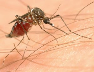
Malaria is a potentially fatal tropical disease that's caused by a parasite known as Plasmodium. It's spread through the bite of an infected female mosquito.
The infected person may have feverish attacks, influenza-like symptoms, tiredness, diarrhoea or a whole range of other symptoms.
Malaria should always be suspected if these symptoms occur within the first year of return from an infected area, and a test should be carried out to exclude the possibility of malaria as soon as possible.
Malaria is one of the leading causes of disease and death in the world. It is estimated that there are 300 to 500 million new cases every year, with 1.5 to 2.7 million deaths worldwide.
Malaria occurs extensively in tropical and subtropical regions.
It used to exist in the UK but fortunately no longer does.
In recent years, about 1,500 people have returned to Britain with malaria that they have contracted abroad - and, of these, an average of 12 die. For this reason it's important to prevent malaria in those travelling to and from the tropics.


What causes malaria?
The malaria parasite, Plasmodium, is a small, single-cell organism (protozoan) that lives as a parasite in man and a specific species of mosquito (Anopheles).
There are four different types of malaria parasite: Plasmodium falciparum is the cause of fatal malaria, while Plasmodium vivax, Plasmodium ovale and Plasmodium malariae cause more benign types of malaria. Falciparum malaria can kill, but the other forms are much less likely to prove fatal.
There are several stages in the life cycle of the parasite, and by and large these are the same for all four types.


How do you catch malaria?
Malaria is passed on by the female Anopheles mosquito biting a person who has malaria parasites in their blood.
The parasites develop in the intestine and salivary glands of the mosquito and can be passed on to other people the next time the mosquito bites.
In man, the parasite travels to the liver via the blood and then out into the bloodstream again, where it invades the red blood corpuscles (the cells which carry oxygen in the blood).
Malaria can also be passed on by blood transfusions and the use of infected needles.
Where does malaria occur?
Malaria occurs where the Anopheles mosquito lives - ie particularly in hot, humid climates.
Plasmodium falciparum is by far the most important malaria parasite in Africa.
There are also areas in: Latin America, Asia, and Oceania, where fatal malaria still occurs.
Plasmodium vivax is the most common in Asia and Latin America, including Central America.
What are the symptoms of the disease?
Normally, 10 to 15 days go by between being infected and the onset of the disease, but it may be longer if the patient has taken a preventive medicine.
On a purely practical level, the most fatal (Plasmodium falciparum) cases develop within three months of leaving the malaria region, while the forms transmitted by Plasmodium vivax and Plasmodium ovale have been recorded to appear up to 22 months later.
Malaria malariae (a rare, benign form) can survive in man for up to 30 years, luckily without causing much discomfort. This form can also be treated, provided you get the right medication.
The actual attacks of malaria develop when the red blood corpuscles burst, releasing a mass of parasites into the blood. The attacks do not begin until a sufficient number of blood corpuscles have been infected with parasites.


What are the characteristics of a malaria attack?
The attack may be what is called uncomplicated or severe.
Classic symptoms would be:
fever and shivering. The attack begins with fever, with the temperature rising as high as 40ºC and falling again over a period of several hours
a poor general condition, feeling unwell and having headaches like influenza
diarrhoea, nausea and vomiting often occur as well.
When the temperature drops, the patient often sweats profusely and feels much better. Then the same day, or one to two days later, further attacks occur with feeling generally unwell, high temperature and so on.
The attacks diminish in the course of a number of weeks, if the patient develops the ability to resist the malaria parasite. But if proper treatment is given, the fever and parasites can disappear within a few days.
But malaria can feel like mild flu. Tiredness can be the only initial symptom or to make diagnosis even more difficult, just simple diarrhoea.
If a case shifts to severe malaria, the classic symptoms above would be expected with increased drowsiness, leading to coma and associated failure of all the major organ systems.
No-one is ever completely immune to malaria, but the concept of semi or partial immunity exists, in which attacks are less severe and less likely to kill. But the price for this is multiple exposures (which kill many children).
Many people form Africa and India assume they have full or partial immunity to malaria, and these people who visit friends and relatives abroad (VFRs) compromise the largest numbers of imported malaria cases in the UK.
In severe malaria the illness may evolve with a number of complications:
low blood pressure (hypotension)
kidney failure
possible haemorrhage (bleeding)
effects on the liver (eg infectious jaundice)
shock and coma may also develop, and the condition may prove fatal.
Cerebral malaria
Severe falciparum malaria can affect the brain and the rest of the central nervous system. It's characterised by changes in the level of consciousness, convulsions and paralysis.
Blackwater-fever
In severe falciparum malaria a large number of the red blood corpuscles are destroyed. Haemoglobin (the red pigment) from the blood corpuscles is excreted in the urine, which therefore is dark and almost the colour of cola.
Late complications
If someone with a benign form of malaria is untreated, anaemia and an enlarged spleen may develop after days or weeks.


Ability to resist malaria attacks
Partial Immunity to malaria develops very slowly and is quickly lost (some estimate within 6 months of leaving the exposure area).
On average one child dies every 30 seconds from malaria in these countries.
It's important to remember that nationals from malarious areas, who return home for holidays, need the same malaria protection as ordinary travellers because partial immunity develops slowly and is rapidly lost.

What can you do yourself?
There's no risk of catching malaria in the UK. But if you visit tropical and subtropical countries, it's important to investigate the chances of catching malaria.
Because the situation can change rapidly: you should talk to a doctor, travel clinic or pharmacist before planning your trip, both as regards to products for malaria prevention and also for expert advice on avoiding other dangers and diseases.
Prevention of malaria is important. If you travel to a region where malaria is prevalent, you should take preventive medication against the parasite and take whatever steps you can to avoid being bitten.
How is the disease diagnosed?
The symptoms of malaria are similar to those of many other diseases and infections that can cause fever or upset the stomach.
Therefore you should always tell your doctor if you have been abroad, especially if you've been to the tropics in the last 12 months.
The gold standard actual diagnosis is made by detecting the parasite in the blood. This is done using a special product mixed with one to two drops of the patient's blood and spreading it on a microscope slide. This is then stained and examined carefully under a microscope.
But many laboratories in the UK and overseas now use rapid antibody based screening tests.
The examination may have to be repeated if the fever has only just begun or preventive medication is to some extent keeping the numbers of the malaria parasite low.
Treatment
The treatment of malaria normally calls for admission to hospital because it may be falciparum malaria that can have a fatal outcome in only a few days or hours.
Outpatient treatment or, worse still, self-treatment of malaria is something only to be undertaken when no qualified medical help is available, ie if you develop malaria in a remote area.
The same antimalarial agents may be used to treat malaria as to prevent it. But if you have caught malaria in spite of using the correct preventive medication, a different product should be used to combat the possibility of resistant parasites.


WHAT IS DENGUE?


Dengue is a viral disease

It is transmitted by the infective bite of Aedes Aegypti mosquito

Man develops disease after 3-14 days (usually 4-7 days) of being bitten by an infective mosquito

It occurs in two forms: Dengue Fever and Dengue Haemorrhagic Fever(DHF)/Dengue Shock Syndrome(DSS).

· Dengue Fever is a severe, flu-like illness

Dengue Haemorrhagic Fever (DHF)/DSS is a more severe form of disease, which may cause death

· Person suspected of having dengue fever or DHF must see a doctor at once


SIGNS & SYMPTOMS OF DENGUE FEVER
Abrupt onset of high fever

Severe frontal headache

Pain behind the eyes(retero-orbital pain) which worsens with eye movement
Muscle and joint pains

Loss of sense of taste and appetite
Measles-like rash over chest and upper limbs
Nausea and vomiting

Minor hemorrhagic manifestations like petechae, bleeding from nose or gums may occur.

Lymphadenopathy with leukopenia and relative lymphocytosis are common. Thrombocytopenia(platelet count £ 100x103) and raised transaminases occur less frequently.


SIGNS & SYMPTOMS OF DENGUE HAEMORRHAGIC FEVER AND DENGUE SHOCK SYNDROME
Symptoms similar to dengue fever. Or history of recent fever. Illness is often biphasic beginning with fever with symptoms as in dengue. During recovery phase of fever patient’s condition worsens markedly with severe weakness, marked restlessness, facial pallor and often diaphoresis and circumoral cyanosis, severe continuous pain abdomen. Liver may be enlarged. Thrombocytopenia ( platelet count £ 100x103 ) also occurs during this phase.
Haemmorhagic phenomenon are frequent and include positive tourniquet test, petechae, easy bruising, bleeding from venepuncture sites, epistaxis, bleeding from mouth & gums and skin rashes.
Frequent vomiting with or without blood. Bleeding from GI tract is an ominous sign that usually follows a prolonged period of shock. There may be signs of plasma leakage indicated by small pleural effusion or ascites. Hepatomegaly is common but is not accompanied by jaundice.
Patient may go into shock manifested by :Pale, cold or clammy skin,sleepiness and restlessness,patient feels thirsty and mouth becomes dry,rapid weak pulse and difficulty in breathing.
CLINICAL AND LABORATORY DIAGNOSIS & CASE DEFINITIONS

DENGUE FEVER:

Suspect case: Acute onset and high fever of 2-7 days duration, and two or more of the following:

Headache,retero-orbital pain, myalgia, arthralgia, rash, hemorrhagic manifestations, and leucopenia.

Probable case: Suspect case and one or more of the following:

Occurance of confirmed cases of dengue in the same place and time. Detection of IgM antibody. IgM antibody indicates current or recent infection and is detectable 6-7 days after onset of illness. If available Mc- Elisa test is more specific.

Confirmed case: Suspect or probable case and one or more of the following:

Isolation of virus or detection of viral genomic sequences. fourfold rise in titres of IgG or IgM antibody. For this at least 2 samples are to be taken- one at the time at the time of reporting to a clinic or a hospital and second shortly before discharge . The optimum interval between two samples should be 10 days. Although serological tests are simpler, they can give false positive results due to cross reaction between antibodies against dengue and other flaviviruses. Confirmatory tests are not necessary for management of cases and should be done to confirm the aetiology of the outbreak.

DENGUE HAEMMORHAGIC FEVER

· Probable or confirmed case of dengue, and

· Haemorrhagic tendencies as described under DHF.

Thrombocytopenia(platelet count £ 100x103 ).Evidence of plasma leakage due to increased vascular permeability, manifested one or more of the following: a rise in average haematocrit for age and sex ³20%, a ³20% drop in haematocrit following volume replacement compared to baseline, signs of plasma leakage indicated by pleural effusion or ascites ( demonstrated by ultrasonography or x-ray), hypoproteinemia. Slight elevation of liver enzymes, hypoproteinemia and low levels of C 3 comlement proteins are commonly observed. Prothrombin, partial thromboplastin, thrombin times may be prolonged in many cases. While a normal WBC count or leukopenia with neutrophils predominating is common initially, a relative lymphocytosis with more than 15% atypical lymphocytes is common when fever subsides.

DENGUE SHOCK SYNDROME

All the criteria for DHF, and

Evidence of circulatory failure as detailed under DSS


MANAGEMENT OF DENGUE FEVER

Early reporting of the suspected dengue fever

Management of dengue fever is symptomatic & supportive.give paracetamol but NO aspirin or brufen. Keep temp. below 390 C. In cases with severe painanalgesics or mild sedatives are to be given. Bed rest is essntial. Oral fluids and electrolyte therapy are required for patients with excessive sweating or vomiting.Follow up for any change in platelet/haematocrit. During afebrile phase(2-3 days after febrile period) check platelet/haematocrit. In convalescent phase no special instructions. Normal diet. Patients almost always recover but often have prolonged asthenia and depression.

MANAGEMENT OF DHF GRADE I & II
Duration is 2-3 days after the febrile phase. Treat on OPD/ inpatient basis. Give ORS. Check platelet/haematocrit.If Hct ³20% start IV therapy. Monitor vitals, urine output, haematocrit .

MANAGEMENT OF DHF GRADE III & IV
Duration is 2-3 days after the febrile phase. Check platelet/haematocrit. Start IV therapy. Monitor vitals, urine output, haematocrit
If Hct is increasing change IV fluid to colloidal solution preferably dextran or plasma. If Hct is decreasing from initial value, give fresh whole blood transfusion. In case of profound give IV fluid bolus one or two times. Give oxygen therapy.
EPIDEMIOLOGY

DISTRIBUTION OF DENGUE/DHF IN INDIA

Disease is prevalent throughout India in most of the metropolitan cities and towns
Outbreaks have also been reported from rural areas of Haryana, Maharashtra & Karnataka

MAGNITUDE OF THE PROBLEM

During 1996 a severe outbreak of Dengue/DHF occurred in Delhi wherein about 10252 cases and 423 deaths were reported

Till date, more then 80 outbreaks have been reported from 16 States/UTs

CAUSATIVE AGENT
It is caused by flaviviruse which has four serotypes; DEN1, DEN2, DEN3, and DEN4. Infection with one serotype provides life long immunity against that serotype but not against other serotypes. Thus people may aquire multiple dengue infections. When there are circulating antibodies against one serotype due to earlier infection and later there is infection with another serotype, it may result in dengue haemorrhagic fever / dengue shock syndrome.

PERIOD OF COMMUNICABILITY
Infected person with Dengue becomes infective to mosquitoes 6 to 12 hours before the onset of the disease and remains so upto 3 to 5 days.
AGE & SEX GROUP AFFECTED

All age groups & both sexes are affected

Deaths are more in children during DHF outbreak

VECTOR OF DENGUE/DENGUE HAEMORRHAGIC FEVER

Aedes aegypti is the principal vector of dengue / dengue haemorrhagic fever. Aedes albopictus also transmits the disease.. .

It is a small, black mosquito with white stripes and is approximately 5 mm in size.

· It takes about 7 to 8 days to develop the virus in its body(extrinsic incubation period) and transmit the disease.

Feeding Habit

Day biter with increased biting activity 2 hours after sunrise and several hours before sunset.

Mainly feeds on human beings in domestic and peridomestic situations

Bites repeatedly

Resting Habit
Rests in the domestic and peridomestic situations
Rests in the dark corners of the houses, on hanging objects like clothes, umbrella, etc. or under the furniture
BREEDING HABITS

Aedes aegypti mosquito breeds in any type of man made containers or storage containers having even a small quantity of water

Eggs of Aedes aegypti can live without water for more then one year

FAVOURED BREEDING PLACES
Desert coolers, Drums, Jars, Pots, Buckets, Flower vases, Plant saucers, Tanks, Cisterns, Bottles, Tins, Tyres, Roof gutters, Refrigerator drip pans, Cement blocks, Cemetery urns, Bamboo stumps, Coconut shells, Tree holes and many more places where rainwater collects or is stored.


CONTROL OF DENGUE/ DENGUE HAEMORRHAGIC FEVER
· Report suspected/probable cases to the health authority, eg. Zonal Health Office of MCD.
No drug or vaccine is available for the treatment of Dengue/DHF. A tetravalent vaccine is under develoment and is undergoing phase I & II trials.

The control of Aedes Aegypti mosquito is the only method of choice

With early detection and proper case management and symptomatic treatment, mortality can be reduced substantially

VECTOR CONTROL MEASURES
1.PERSONAL PROPHYLATIC MEASURES

· Use of mosquito repellent creams, liquids, coils, mats etc.

· Wearing of full sleeve shirts and full pants with socks

· Use of bednets for sleeping infants and young children during day time to prevent mosquito bite

2. VECTOR CONTROL

· As Aedes aegypti breeds in containers and receptacles detection & elimination of mosquito breeding sources is the most important activity.
· Management of roof tops, porticos and sunshades
· Proper covering of stored water
· Reliable water supply
· Observation of weekly dry day
5. HEALTH EDUCATION AND COMMUNITY PARTICIPATION

Impart knowledge to common people regarding the disease and vector through various media sources like T.v., Radio, Cinema slides, etc. Sensitilizing and involving the community for detection of Aedes breeding places and their elimination.

DO’S AND DON’TS

Remove water from coolers and other small containers at least once in a week

Use aerosol during day time to prevent the bites of mosquitoes

Do not wear clothes that expose arms and legs
Children should not be allowed to play in shorts and half sleeved clothes
Use mosquito nets or mosquito repellents while sleeping during day time

EARLY WARNING SIGNALS FOR DF/DHF OUTBREAK

Sudden increase in reporting of suspected cases with clutering in time and place and fitting into endemicity/ seasonality of disease.

Enhanced vector density as indicated through household larvae index/ container index / Breteau index with reference to vector mosquito.

Detection of viral activity either in vector or man.

. .


FREQUENTLY ASKED QUESTIONS

Q1. What is the difference between DF and DHF?

Ans. DHF resembles DF during initial phase of illness. However, thrombocytopenia accompanied by / followed by a rising haematocrit distinguishes DHF from DF and other diseases in the differential diagnosis, viz. malaria, leptospirosis, other arthropod born viral infections, influenza, meningococcemia and bacterial shock.


Q2. Do haemorrhagic manifestations occur only in DHF?

Ans. No. Minor haemorrhagic manifestations occur in some of the DF patients also.These cases have to be differentiated from DHF.

Q3. Why do some people have DF and others have DHF, although same flaviviruses cause both?

Ans. It is not clear with certainity why does DHF occur in some cases when the most have only dengue fever. It .is suggested that when there are circulating antibodies against one serotype due to earlier infection and later there is infection with another serotype, it may result in dengue haemorrhagic fever / dengue shock syndrome. The risk of DHF is 0.2% during the first dengue infection but it increases 10-fold during infection with a second serotype of dengue virus. However, the circulation of multiple dengue serotypes does not always produce DHF.

Q4. What is the difference in management of DF and DHF?

Ans. The manifestations and management of DF and DHF during febrile phase are the same. It is during afebrile phase of 2-3 days (critical phase) following the febrile phase that if plasma leakage occurs, it has to be managed by IV fluids ( ringer lactate/ normal saline ) in most cases. This phase lasts for 24-48 hours.

Q5. What is the role of platelet transfusion in cases of thrombocytopenia?

Ans. In most cases of DHF/DSS IV fluids / plasma expanders are adequate. However blood transfusion is indicated when haematocrit falls due to severe bleeding ( refractory shock ) and not due to fluid therapy. Fresh whole blood is preferable and volume administered should be just adequate to raise the RBC concentration to normal.
Fresh frozen plasma and / or concentrated platelets may be indicated in a few cases when disseminated intravascular coagulation (DIC) causes massive bleeding.

Q6. What is the role of insecticide spray in control of an outbreak of DF/DHF?

Ans. Residual insecticide spray in the household where a case of DF/DHF is reported and its neighbourhood kills the infected mosquitoes and thus prevents its further spread. However, generalized use of insecticides as space spray or ultra low volume spray is only of a limited value as these reduce the mosquito population only for a short period. In fact they lead to a false sense of security. Therefore, they are used as a supplement to source reduction and anti-larval measures which produce a lasting effect.

Saturday, July 3, 2010

What is Yoga?


Introduction to Yoga


Yoga is a path towards total harmony of body, mind, and spirit.

The word Yoga comes from the Sanskrit word yuj, which means union. Union of the individual consciousness with the universal consciousness.

Yoga is not merely a form of exercise for the body. It is an ancient wisdom - for a healthier, happier, and more peaceful way of living - which ultimately leads to union with the Self.

It is an inherent desire in humans to be happy. The ancient sages, through inquiry about life, were able to reach a state of consciousness in which the secrets of healthier, happier, and meaningful living were revealed to them.

Though yoga comes from Hinduism, the knowledge of yoga transcends any religion or culture. Its application is universal!
Benefits of Yoga

introduction to yoga

* Helps in releasing toxins from the body
* Channelises energy flow
* Improves the flexibility of muscles, range of motions of joints.
* Corrects the posture and alignment of the body
* Regulates the digestive, endocrine, and circulatory systems
* Strengthens and revitalizes the internal organs, leading to a healthy and youthful body
* Helps to lose weight, cure asthma, diabetes, heart problems, and many chronic diseases


The practice of Yoga benefits the body, mind, and spirit. It is accessible to everyone. While some of these benefits can be expressed in words, others are beyond description, and can only be experienced.
Benefits
Benefits of yoga

Regular practice of yoga, benefits the body in the following ways

* Yoga improves flexibility of muscles and joints
* Yoga strengthens muscles and bones
* Yoga improves posture and body alignment
* Yoga improves digestion, circulation, and immunity
* Yoga enhances function of neurological and endocrine organs
* Yoga prevents and provides relief from chronic illnesses, such as chronic pain syndromes,anxiety and panic disorders, depression, sleep disorders, chronic fatigue syndrome, and high blood pressure.
* Overall the body feels healthier, more energetic. It is less prone to diseases and effects of day-to-day stress.


Yoga and Spirituality

Spirituality is a harmonious blend of outer silence and inner celebration and also inner silence and outer celebration.
Inner Silence and Outer Celebration

Anytime you are confused, your mind is in conflict, do asanas [Yoga postures], sit in asana. You will see right away that clarity dawns. Effect of asana is to clear out all conflicts, duality. The human tendency is to cling to the negative. However, the technology called yoga and meditation helps the mind to effortlessly transcend the pessimism and negativity and truly experience the exhilarating life force.
Spirituality and Yoga

For the body we have asanas. For the mind we have pranayamas which provides emotional and mental well-being. Asanas also work on the mind and pranayamas on the body -- there are no strict boundaries. Meditation removes spiritual misery, dejection, darkness, and depression. That is why yoga, Yogas-chitta- vritti-nirodhaha, stops the chitta-vritti, or the endless activity of the chitta.

In order to make life more beautiful. Heyam dukham anahatam - to stop misery from entering our world- in the physical, mental, emotional, and spiritual spheres - we do Yoga.

We need to do a cleansing process within ourselves. In sleep we get rid of fatigue, but the deeper stresses remain in our body. Some meditation, yoga, and Sudarshan Kriya cleanse the whole system. You blossom from inside and become centered. Otherwise, our peace is disturbed by small things.

When attention is given to the spiritual aspect of one’s life, it brings responsibility, a sense of belongingness, and compassion and caring for the whole of humanity.

The Knowledge that unites love and wisdom, that uplifts the spirit is spirituality. The Knowledge which gives you a broad vision and a big heart is Spirituality.


In Depth of Yoga

Have you imagined delving into the depth of your existence? How would it be?
Why should one understand the depths of yoga?

We plan our vacations with great detail – mode of travel, selection of place, sightseeing tours. We do have a good time, but how are we at the end of the journey – tired, exhausted? Why is this so?
In Depth of Yoga

Our senses have limited capacity to enjoy, but our mind has an infinite desire for joy. The joy, which comes through the five senses, is limited.

Most of us think that the joy is in objects, but it is inside us in the senses, not out in the objects. Each of the senses leads you to a point inside you, which is a fountain of joy, deep within you.

Clinging on to sense objects make you miserable after a while. Move on to the experience of that pleasure - from the object of pleasure. From the object move to the Center, move to that which experiences the pleasure.
Path of Yoga - the inner compass

How do we understand yoga in depth? The path of Yoga leads us inward, to experience the union with the True Self. The purpose of creation is to go from two to one. The ancient Rishis called it Yoga.

Our breath plays a very important role. The breath is the connecting link between the inner world of the mind and the outer world of body and environment.

There are four inner modes of consciousness

* Mind
* Intellect
* Memory
* Ego

When the mind is not centered there is restlessness. Yoga is the restraint of thought waves, Vritis. Modulations in the mind are called Vritis. Subsiding these waves in the mind is called Yoga. When thoughts are constrained, then there is communion with the Lord, Divinity, and the Cosmic Being.

Vritis are like a whirlpool in the pond of mind. When mind and memory get excited, thought waves arise in you. When excitement in mind is calmed, the Vritis too gets silenced. Only when the whirlpool calms down, then can you see the real depths of the ocean. The wave uniting with this depth is Yoga.
Move to the Center – with proper guidance and practice

Learn Yoga under a Guru. You cannot operate on your own body even if you are a surgeon. To move around in a small new place, you need a guide to show you way. Then to walk into a completely unknown realm like consciousness, you certainly need guidance. A lit candle can light million more candles, but a matchbox even though having a potential, cannot light up on its own.

Guru itself means something that which is enormously big, immeasurable. Guru is someone so vast but still tangible and available. Guru is the infinite love yet in a bodily form.
The practice

The Yoga seeker (sadhak) gains depth with proper guidance and with the proper practice of Yoga. Yoga asanas, pranayamas, and meditation, enable us to deepen our sadhana (practice)

* The asanas help to center the mind and body
* The breath is a bridge, connecting with the energy manifesting in the body
* The pranayama energises and calms the system

Thus by refining our practice, thoughts diminish enabling us to go deep in Meditation. When we can clearly see through the levels of existence, we then attain a state of Yoga, oneness with the universe.

What is Lung Cancer ?


LUNG CANCER
Cancer is a class of diseases characterized by out-of-control cell growth, and lung cancer occurs when this uncontrolled cell growth begins in one or both lungs. Rather than developing into healthy, normal lung tissue, these abnormal cells continue dividing and form lumps or masses of tissue called tumors. Tumors interfere with the main function of the lung, which is to provide the bloodstream with oxygen to be carried to the entire body. If a tumor stays in one spot and demonstrates limited growth, it is generally considered to be benign.
doctors viewing a lung x-ray

More dangerous, or malignant, tumors form when the cancer cells migrate to other parts of the body through the blood or lymph system. When a tumor successfully spreads to other parts of the body and grows, invading and destroying other healthy tissues, it is said to have metastasized. This process itself is called metastasis, and the result is a more serious condition that is very difficult to treat.

Lung cancer is called "primary" if the cancer originates in the lungs and "secondary" if it originates elsewhere in the body but has metastasized to the lungs. These two types are considered different cancers from diagnostic and treatment perspectives.

In 2007, about 15% of all cancer diagnoses and 29% of all cancer deaths were due to lung cancer. It is the number one cause of death from cancer every year and the second most diagnosed after breast and prostate cancers (for women and men, respectively). Lung cancer is usually found in older persons because it develops over a long period of time.
How is lung cancer classified?

Lung cancer can be broadly classified into two main types based on the cancer's appearance under a microscope: non-small cell lung cancer and small cell lung cancer. Non-small cell lung cancer (NSCLC) accounts for 80% of lung cancers, while small cell lung cancer accounts for the remaining 20%.

NSCLC can be further divided into four different types, each with different treatment options:

* Squamous cell carcinoma or epidermoid carcinoma. As the most common type of NSCLC and the most common type of lung cancer in men, squamous cell carcinoma forms in the lining of the bronchial tubes.
* Adenocarcinoma. As the most common type of lung cancer in women and in nonsmokers, adenocarcinoma forms in the mucus-producing glands of the lungs.
* Bronchioalveolar carcinoma. This type of lung cancer is a rare type of adenocarcinoma that forms near the lungs' air sacs.
* Large-cell undifferentiated carcinoma. A rapidly growing cancer, large-cell undifferentiated carcinomas form near the outer edges or surface of the lungs.

Small cell lung cancer (SCLC) is characterized by small cells that multiply quickly and form large tumors that travel throughout the body. Almost all cases of SCLC are due to smoking.

What causes cancer?
Cancer is ultimately the result of cells that uncontrollably grow and do not die. Normal cells in the body follow an orderly path of growth, division, and death. Programmed cell death is called apoptosis, and when this process breaks down, cancer begins to form. Unlike regular cells, cancer cells do not experience programmatic death and instead continue to grow and divide. This leads to a mass of abnormal cells that grows out of control.

Lung cancer occurs when a lung cell's gene mutation makes the cell unable to correct DNA damage and unable to commit suicide. Mutations can occur for a variety of reasons. Most lung cancers are the result of inhaling carcinogenic substances.
Carcinogens

Carcinogens are a class of substances that are directly responsible for damaging DNA, promoting or aiding cancer. Tobacco, asbestos, arsenic, radiation such as gamma and x-rays, the sun, and compounds in car exhaust fumes are all examples of carcinogens. When our bodies are exposed to carcinogens, free radicals are formed that try to steal electrons from other molecules in the body. These free radicals damage cells and affect their ability to function and divide normally.

About 87% of lung cancers are related to smoking and inhaling the carcinogens in tobacco smoke. Even exposure to second-hand smoke can damage cells so that cancer forms.
Genes

Cancer can be the result of a genetic predisposition that is inherited from family members. It is possible to be born with certain genetic mutations or a fault in a gene that makes one statistically more likely to develop cancer later in life. Genetic predispositions are thought to either directly cause lung cancer or greatly increase one's chances of developing lung cancer from exposure to certain environmental factors.


What are the symptoms of lung cancer?

Cancer symptoms are quite varied and depend on where the cancer is located, where it has spread, and how big the tumor is. Lung cancer symptoms may take years before appearing, usually after the disease is in an advanced stage.

Many symptoms of lung cancer affect the chest and air passages. These include:

* Persistent or intense coughing
* Pain in the chest shoulder, or back from coughing
* Changes in color of the mucus that is coughed up from the lower airways (sputum)
* Difficulty breathing and swallowing
* Hoarseness of the voice
* Harsh sounds while breathing (stridor)
* Chronic bronchitis or pneumonia
* Coughing up blood, or blood in the sputum

If the lung cancer spreads, or metastasizes, additional symptoms can present themselves in the newly affected area. Swollen or enlarged lymph nodes are common and likely to be present early. If cancer spreads to the brain, patients may experience vertigo, headaches, or seizures. In addition, the liver may become enlarged and cause jaundice and bones can become painful, brittle, and broken. It is also possible for the cancer to infect the adrenal glands resulting in hormone level changes.

As lung cancer cells spread and use more of the body's energy, it is possible to present symptoms that may also be associated with many other ailments. These include:

* Fever
* Fatigue
* Unexplained weight loss
* Pain in joints or bones
* Problems with brain function and memory
* Swelling in the neck or face
* General weakness
* Bleeding and blood clots

How is lung cancer diagnosed and staged?
examination

Physicians use information revealed by symptoms as well as several other procedures in order to diagnose lung cancer. Common imaging techniques include chest X-rays, bronchoscopy (a thin tube with a camera on one end), CT scans, MRI scans, and PET scans. Physicians will also conduct a physical examination, a chest examination, and an analysis of blood in the sputum. All of these procedures are designed to detect where the tumor is located and what additional organs may be affected by it.

Although the above diagnostic techniques provided important information, extracting cancer cells and looking at them under a microscope is the only absolute way to diagnose lung cancer. This procedure is called a biopsy. If the biopsy confirms lung cancer, a pathologist will determine whether it is non-small cell lung cancer or small cell lung cancer.

After a diagnosis is made, an oncologist will determine the stage of the cancer by finding out how far the cancer has spread. The stage determines which choices will be available for treatment and informs prognosis. The most common cancer staging method is called the TNM system. T (1-4) indicates the size and direct extent of the primary tumor, N (0-3) indicates the degree to which the cancer has spread to nearby lymph nodes, and M (0-1) indicates whether the cancer has metastasized to other organs in the body. A small tumor that has not spread to lymph nodes or distant organs may be staged as (T1, N0, M0), for example.

For non-small cell lung cancer, TNM descriptions lead to a simpler categorization of stages. These stages are labeled from I to IV, where lower numbers indicate earlier stages where the cancer has spread less. More specifically:

* Stage I is when the tumor is found only in one lung and in no lymph nodes.
* Stage II is when the cancer has spread to the lymph nodes surrounding the infected lung.
* Stage IIIa is when the cancer has spread to lymph nodes around the trachea, chest wall, and diaphragm, on the same side as the infected lung.
* Stage IIIb is when the cancer has spread to lymph nodes on the other lung or in the neck.
* Stage IV is when the cancer has spread throughout the rest of the body and other parts of the lungs.

Small cell lung cancer has two stages: limited or extensive. In the limited stage, the tumor exists in one lung and in nearby lymph nodes. In the extensive stage, the tumor has infected the other lung as well as other organs in the body.
How is lung cancer treated?

Lung cancer treatments depend on the type of cancer, the stage of the cancer (how much it has spread), age, health status, and additional personal characteristics. As there is usually no single treatment for cancer, patients often receive a combination of therapies and palliative care. The main lung cancer treatments are surgery, chemotherapy, and/or radiation. However, there also have been recent developments in the fields of immunotherapy, hormone therapy, and gene therapy.
Surgery

Surgery is the oldest known treatment for cancer. If a cancer is in stage I or II and has not metastasized, it is possible to completely cure a patient by surgically removing the tumor and the nearby lymph nodes. After the disease has spread, however, it is nearly impossible to remove all of the cancer cells.
surgery room

Lung cancer surgery is performed by a specially trained thoracic surgeon. After removing the tumor and the surrounding margin of tissue, the margin is further studied to see if cancer cells are present. If no cancer is found in the tissue surrounding the tumor, it is considered a "negative margin." A "positive margin" may require the surgeon to remove more of the lung tissue.

Lung cancer surgery can be curative or palliative. Curative surgery aims to cure a patient with early stage lung cancer by removing all of the cancerous tissue. Palliative surgery aims to remove an obstruction or open an airway, making the patient more comfortable but not necessarily removing the cancer.

Surgery carries side effects - most notably pain and infection. Lung cancer surgery is an invasive procedure that can cause harm to the surrounding body parts. Doctors will usually provide several options for alleviating any pain from surgery. Antibiotics are commonly used to prevent infections that may occur at the site of the wound or elsewhere inside the body.
Radiation

Radiation treatment, also known as radiotherapy, destroys or shrinks lung cancer tumors by focusing high-energy rays on the cancer cells. This causes damage to the molecules that make up the cancer cells and leads them to commit suicide. Radiotherapy utilizes high-energy gamma-rays that are emitted from metals such as radium or high-energy x-rays that are created in a special machine. Radiation can be used as the main treatment for lung cancer, to kill remaining cells after surgery, or to kill cancer cells that have metastasized.
Chemotherapy

Chemotherapy utilizes strong chemicals that interfere with the cell division process - damaging proteins or DNA - so that cancer cells will commit suicide. These treatments target any rapidly dividing cells (not just cancer cells), but normal cells usually can recover from any chemical-induced damage while cancer cells cannot. Chemotherapy is considered systemic because its medicines travel throughout the entire body, killing the original tumor cells as well as cancer cells that have spread throughout the body.

A medical oncologist will usually prescribe chemotherapy drugs for lung cancer to be taken intravenously, but there are also drugs available in tablet, capsule, and liquid form. Chemotherapy treatment occurs in cycles so the body has time to heal between doses, and dosages are determined by the type of lung cancer, the type of drug, and how the person responds to treatment. Medicines may be administered daily, weekly, or monthly, and can continue for months or even years.

Combination therapies often include multiple types of chemotherapy, and chemotherapy is also given as adjuvant therapy as a complement to surgery and radiation. Adjuvant therapy is designed to reduce the risk of cancer recurrence after surgery and killing any cancer cells that exist after surgery. Chemotherapy can be given before surgery, called neo-adjuvant therapy, to shrink tumors and to make surgery more successful.

Chemotherapy carries several common side effects, but they depend on the type of chemotherapy and the health of the patient. These include nausea and vomiting, appetite loss, diarrhea, hair loss, fatigue from anemia, infections, bleeding, and mouth sores. Many of these side effects are only temporarily felt during treatment, and several drugs exist to help patients cope with the symptoms.
Other Treatments

Researchers continue to search for ways to improve lung cancer treatments and find new methods of treating the disease. Targeted therapies are designed to only treat cancer cells while leaving alone normal and healthy lung cells. These include monoclonal antibodies that travel directly to the cancer cells and release drugs or radiation, anti-angiogenesis agents that interfere with the blood supply creation mechanism of cancer cells, and growth factor inhibitors that block the effects of growth factors and disallow the cancerous cells to grow. There is also some research in the area of lung cancer vaccines that first transform cancer cells so they are no longer cancerous. However, the cells will exist such that the body's immune system can recognize the cancerous cells as foreign and attack them. These targeted therapies are also called immunotherapies because the treatment tweaks the body's natural immune responses.
How can lung cancer be prevented?

Cancers that are closely linked to certain behaviors are the easiest to prevent. For example, choosing not to smoke tobacco or drink alcohol significantly lowers the risk of several types of cancer - most notably lung, throat, mouth, and liver cancer. Even if you are a current tobacco user, quitting can still greatly reduce your chances of getting cancer. The most important preventive measure you can take to avoid lung cancer is to quit smoking.

Quitting smoking will also reduce your risk of several other types of cancer including esophagus, pancreas, larynx, and bladder cancer. If you quit smoking, you will usually reap additional benefits such as lower blood pressure, enhanced blood circulation, and increased lung capacity.

Exposure to tobacco smoke is not the only risk factor for lung cancer though. Those who have come into contact with asbestos, radon, and secondhand smoke also have an increased risk of developing lung cancer. In addition, having a family member who developed lung cancer without being exposed to carcinogens could mean that you have a genetic predisposition for developing the disease, increasing your overall risk.

Screening techniques are designed to find cancer at the earliest stage so that the most treatment options are available, increasing survival rates and avoiding highly invasive procedures. Most lung cancers are detected in the late stages of the disease after they have spread and are harder to treat. Although there currently do not exist approved screening tests for lung cancer that improve survival or detect localized disease, there is promising research underway. Advocates of screening recommend that certain high risk groups be screened. This includes persons age 60 or older with a history of smoking, previous lung tumors, or chronic obstructive pulmonary disease (COPD). Possible lung cancer screening tests include analysis of sputum cells, fiberoptic examination of bronchial passages (bronchoscopy), and low-dose spiral CT scans.